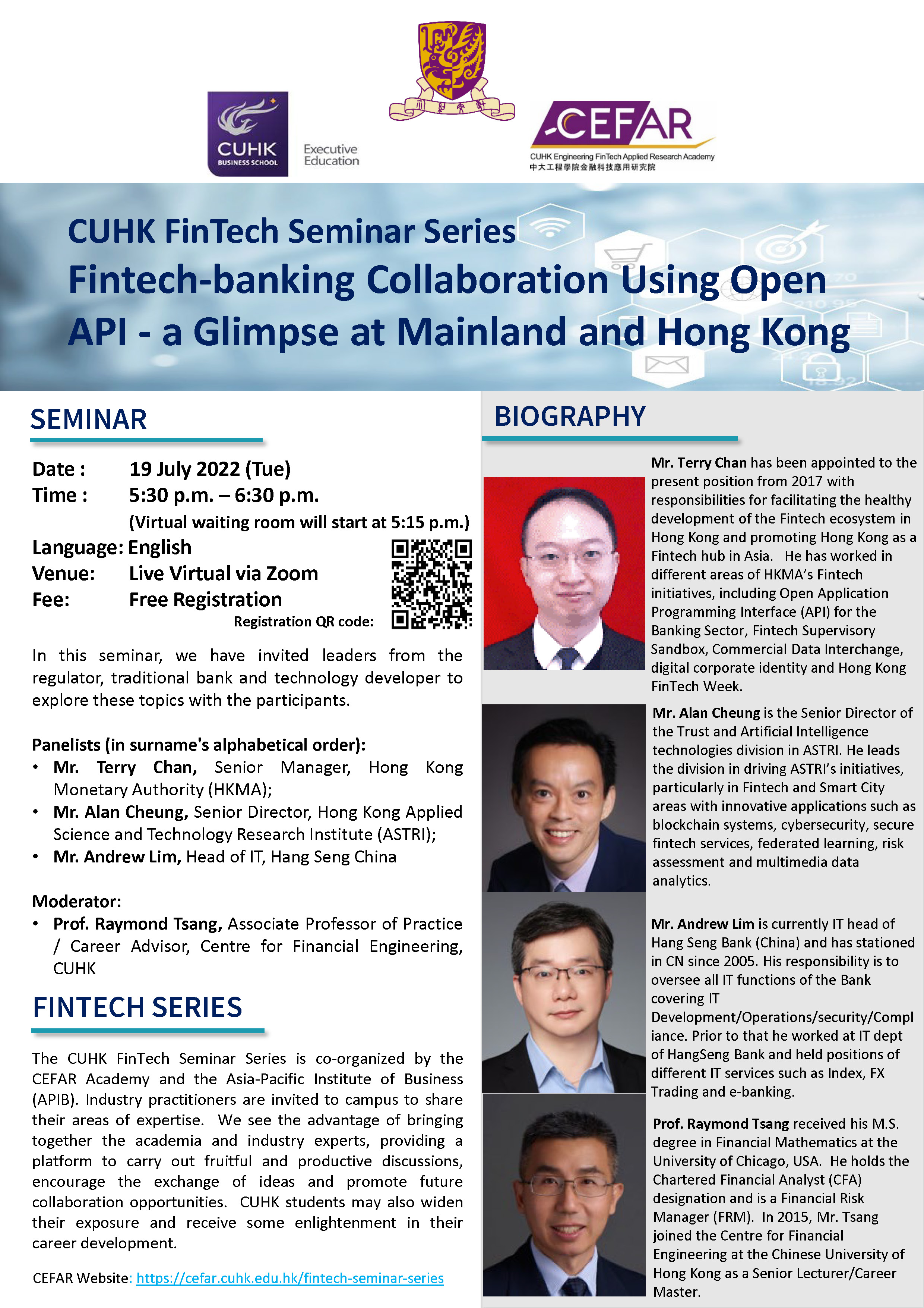
中大AI 「驗毒」 40毫秒即知

香港中文大學昨日表示,中大研究團隊開發了一個人工智能系統(AI),可快速自動檢測胸部電腦斷層掃描(CT)影像上的新冠病毒徵狀,準確度高達96%,料可用於診斷、監察治療病情進展及預測治療成效。
 
中大醫學院影像及介入放射學系助理教授蘇宛彤表示,AI具有明顯的速度優勢。傳統臨床閱片流程上,醫生檢查一個CT影像需時約5分鐘至10分鐘,而AI在40毫秒內即可完成,可提高臨床診斷效率及減省相關人手。中大醫學院影像及介入放射學系系主任余俊豪則指,現時AI主要收集未變異的病毒數據,相信如未來再有變種病毒相關確診個案,可加入AI系統內
Date: 
Thursday, April 22, 2021
Media: 
文匯報

AI分析新冠患者CT影像 僅0.04秒揪出肺部病灶

香港中文大學研究團隊開發了一個人工智能(AI)系統,可快速及準確地自動檢測胸部電腦斷層掃描(CT)影像上的新冠肺炎感染病灶,為臨牀醫生提供即時可靠的診斷結果,而系統亦僅需四十毫秒,即百分之四秒內即可準確評估整個三維CT影像,較傳統的臨牀閱片流程需時五至十分鐘更具效率。該項研究近期已發表在「Nature」旗下綜合期刊《npj Digital Medicine》。

Date: 
Thursday, April 22, 2021
Media: 
Sing Tao Daily

CUHK Research Team Develops an AI System for Detecting COVID-19 Infections in CT with a Privacy Preserving Multinational Validation Study

Date: 
2021-04-21
Thumbnail: 
Body: 
A multidisciplinary research team from CUHK has developed an artificial intelligence (AI) system for the automated, rapid and accurate detection of COVID-19 infections in chest computed tomography (CT) images. The team was led by Professor Qi DOU and Professor Pheng Ann HENG from the Department of Computer Science and Engineering, Faculty of Engineering, Dr. Tiffany SO and Professor Simon YU from the Department of Imaging and Interventional Radiology, Faculty of Medicine. 
 
Using new federated learning techniques, the AI system is trained on multicentre data in Hong Kong without the need to centralise data in one place, thus protecting patient privacy. “The established AI system is validated on multiple, unseen, independent external cohorts from mainland China and Europe, showing the potential and feasibility to build large-scale medical datasets with privacy protection, so as to rapidly develop reliable AI models amidst a global disease outbreak such as the COVID-19 pandemic,” said Professor Qi DOU from the Department of Computer Science and Engineering. A recent research article describing the outcomes from the study has been published in the npj Digital Medicine, part of the Nature Partner Journals series. 
 
Accurate COVID-19 CT lesion detection with federated deep learning
 
COVID-19 has presented a public health crisis worldwide. In Hong Kong, even with advanced healthcare service systems, the rapidly evolving COVID-19 pandemic has been overwhelming the existing clinical systems and burdening frontline radiologists with an unprecedentedly large emergency workload for data analysis and medical image interpretation. Given this situation, automated diagnostic methods with AI are extremely helpful to facilitate effective management of COVID-19. Radiological imaging can play a complementary role together with clinical diagnostic testing in COVID-19 diagnosis, and can effectively assess the severity and progression in the course of disease. The team in close collaboration with engineering and clinical experts has developed an accurate AI system for automated detection of COVID-19 lesions from CT images, which can provide immediately available results, alleviating the burden of clinicians in interpreting images. Professor Pheng Ann HENG from the Department of Computer Science and Engineering said, “Making use of the cutting-edge federated learning techniques, the AI system can effectively coordinate the patient data across multiple clinical centres in Hong Kong, including Prince of Wales Hospital, for model development. Given the unavoidable challenge of data heterogeneity in medical images, multicentre collaborative effort is essential to capture diverse data distributions, which enhances model reliability for unleashing the potential of AI-powered medical image diagnosis in complex clinical practice.”
 
Model robustness and generalisability on multi-national validation cohorts
 
The established AI model has been externally validated on multiple unseen cohorts from mainland China and Germany. Experimental outcomes revealed that the AI model yields a competitive performance in lesion detection in comparison with radiologist interpretation of chest CT across local, regional and global patients. This wide validation and applicability on cohorts with various imaging scanners and different demographics show outstanding robustness and generalisability of the established AI model in complex real-world situations. Dr. Tiffany SO, Assistant Professor from the Department of Imaging and Interventional Radiology at the Faculty of Medicine, stated, “Besides a high diagnostic accuracy, the AI system can also present a remarkable speed advantage to clinician interpretation. In traditional clinical diagnosis, review and interpretation of a single chest CT takes at least 5-10 minutes for clinicians. In contrast, the AI system can accurately evaluate the same CT data in around 40 ms, showing immense potential to support real-time clinical practice.”
 
This latest study demonstrates the use of privacy preserving AI in responding to a global disease outbreak. In the rapidly evolving pandemic of COVID-19, there is apparently no time to set up complicated data sharing agreements across institutions or even countries. Professor Simon YU, Professor and Chairman of the Department of Imaging and Interventional Radiology at the Faculty of Medicine, added, “Privacy preserving machine learning acts as an important enabler under such situations to gather efforts on digital medicine technology for providing reliable clinical assistance for timely patient care. This study demonstrates the feasibility and efficacy of federated learning for COVID-19 image analysis, where collaborative effort is especially valuable at a time of global crisis. More importantly, beyond assisting COVID-19 management, we believe that AI, which protects patient privacy and achieves reliable generalisability in practice, has enormous potential to revolutionise smart hospitals and healthcare systems in Hong Kong and worldwide.”

A multidisciplinary research team from CUHK has developed an artificial intelligence (AI) system for the automated, rapid and accurate detection of COVID-19 infections in chest computed tomography (CT) images.The research team includes: Mr. Quande LIU (1st from right), PhD student, and Prof. Qi DOU (2nd from left), Assistant Professor, Department of Computer Science and Engineering; and Dr. Tiffany SO (1st from left), Assistant Professor, and Prof. Simon YU (2nd from right), Chairman, Department of Imaging and Interventional Radiology, Faculty of Medicine; CUHK.

Prof. Simon YU believes that AI, which protects patient privacy and achieves reliable generalisability in practice, has enormous potential to revolutionise smart hospitals and healthcare systems in Hong Kong and worldwide.

Dr. Tiffany SO states that review and interpretation of a single chest CT takes at least 5-10 minutes for clinicians in traditional clinical diagnosis. In contrast, the AI system can accurately evaluate the same CT data in around 40 ms, showing immense potential to support real-time clinical practice. 

Prof. Qi DOU states that the established AI system is validated on multiple independent cohorts, showing the potential and feasibility to build large-scale medical datasets with privacy protection, so as to rapidly develop reliable AI models amidst a global disease outbreak such as the COVID-19 pandemic.


YouTube_news: 
Filter: Dept: 
Faculty
CSE
Media Release

CUHK Engineering Recognised in the International Exhibition of Inventions Geneva

Date: 
2021-04-10
Thumbnail: 
Body: 
Scientific innovations developed by Faculty of Engineering, CUHK received recognition for outstanding performance in the International Exhibition of Inventions Geneva 2021. 
 
Summary of CUHK Engineering award-winning projects:
 
Gold Medal:
Self-powered smart watch and wristband enabled by embedded generator (link to press release)
 
Members: Professor Wei Hsin LIAO, Dr. Mingjing CAI and Dr. Jiahua WANG, from the Department of Mechanical and Automation Engineering, Faculty of Engineering
The limited battery life of smart watches and wristbands remains a pain point. The research team has designed an embedded and compact electromagnetic generator that can self-power wearable gadgets. Unlike existing products, the invention uses a novel magnetic frequency-up converter and harnesses the kinetic energy of human motion. The converter transforms the low-frequency arm swing to achieve the desired output power.
 
Gold Medal:
Highly Sensitive Gas Sensing and Control System (link to press release)
Members: Professor Wei REN, Dr. Ke XU, from the Department of Mechanical and Automation Engineering, Faculty of Engineering
The research team has invented a portable and highly sensitive gas sensing system that can provide a variety of information about the concentration, temperature, and pressure of multiple harmful gas components such as CO, NOx, NH3, SO2 in real time. It has adopted an advanced laser spectroscopy technology and artificial intelligence algorithm allowing direct application to the fields of environmental protection and medical treatment, including exhaust monitoring in power plants, the petrochemical industry and vehicle emission, as well as components monitoring in patient breath.
 
Silver Medal:
Soliste – A Social Listening System for Understanding Your Customer
Members: Professor Kam Fai WONG and Dr. Gabriel FUNG, from the Department of Systems Engineering and Engineering Management, Faculty of Engineering
 
Silver Medal:
Harvesting energy from walking human body (link to press release)
Members: Professor Wei Hsin LIAO, Dr. Fei GAO, Gaoyu LIU, Brendon Lik-Hang CHUNG and Hugo Hung-Tin CHAN, from the Department of Mechanical and Automation Engineering, Faculty of Engineering
 
Bronze Medal:
QuickCAS: An easy-to-use analysis system for quick detection of infectious pathogens in clinical samples (link to press release)
Members: Professor Li ZHANG, Dr Lidong YANG and Wai Shing LIU, from the Department of Mechanical and Automation Engineering, Faculty of Engineering; Professor Joseph SUNG, Emeritus Professor of CUHK; Dr. Sunny WONG, from the Department of Medicine and Therapeutics, Faculty of Medicine; Professor Philip CHIU, Dr Kai Fung CHAN, from the Chow Yuk Ho Technology Centre for Innovative Medicine, Faculty of Medicine; Professor Margaret IP, Department of Microbiology, Faculty of Medicine
 

The embedded energy harvester in smart watch and wristband, developed by the research team led by Professor Wei Hsin LIAO, receives the Gold Medal.

Professor REN’s research team is collaborating with power plant companies in the mainland China to apply the gas sensing and control system.

Professor Liao (right) and his Postdoctoral fellow Gao Fei (left) develop this energy harvester device in six months’ time, and the device is extremely light with only 307 grams.

The first generation of microrobotic detection system, “QuickCAS”, aims at detecting Clostridium difficile (C. diff), a common pathogen of nosocomial infection.


Filter: Dept: 
Faculty
MAE
SEEM
Media Release
Name: 
Ken Long LEE
Title ( post ): 
President
Department: 
Biomedical Engineering Alumni Association of The Chinese University of Hong Kong
email: 
cuhkbmealumni [at] gmail.com
Avatar: 
Class: 
alumni_association
glossary_index: 
L
Name: 
HUANG Chaoran
Title ( post ): 
Assistant Professor
Department: 
Electronic Engineering
email: 
crhuang [at] ee.cuhk.edu.hk
phone: 
3943 8605
website: 
http://www.ee.cuhk.edu.hk/en-gb/people/academic-staff/professors/prof-huang-chaoran
Avatar: 
Class: 
faculty_member
Chinese Name: 
黃超然
glossary_index: 
H

CUHK Develops Biohybrid Soft Microrobots with a Rapid Endoluminal Delivery Strategy for Gastrointestinal (GI) Diseases

Date: 
2021-04-09
Thumbnail: 
Body: 
A collaborative research team led by Professor Li ZHANG from the Faculty of Engineering, Professor Joseph Jao-yiu SUNG and Professor Philip Wai-yan CHIU from the Faculty of Medicine at The Chinese University of Hong Kong (CUHK) has developed biohybrid soft microrobots with an endoscopy-assisted magnetic navigation strategy for rapid endoluminal delivery. This work provides a new enabling technology for medical microrobot-based minimally invasive intervention and has the potential for treating various diseases in tiny and tortuous lumens which are hard-to-reach or inaccessible by regular medical devices.
Endoscopy-assisted magnetic navigation of biohybrid soft microrobots with rapid endoluminal delivery and imaging,
Science Robotics, Vol. 6, Issue 52, eabd2813, 2021
 
 
Medical micro-/nanorobots and their challenges in in vivo applications
 
For medical microrobots to navigate in tiny and tortuous lumens inside the human body, there are several key challenges to be extensively investigated for in vivo applications, including multi-functionalities and safety, adaptivity in a dynamic physiological environment with biological barriers, and real-time imaging and control.
 
Endoscopy-assisted magnetic navigation of biohybrid soft microrobots with rapid endoluminal delivery and imaging
 
The research team has developed stem-cell-based soft and resilient microrobots, named magnetic stem cell spheroid microrobots (MSCSMs), which are mainly composed of stem cells (~98%) and a tiny portion of magnetic particles (~2%). The soft microrobots, which possess an elastic modulus close to the human brain tissue, not only have rapid response and precise targeting capability under the magnetic field, but also show excellent adaptability to the complex surroundings by self-alternating the shape during navigation inside the body. The stem cells can be harvested from the host so as to minimise immune responses during the in vivo delivery. In addition, the soft microrobots are capable of real-time in vivo tracking by various clinical imaging techniques, including endoscopy and ultrasound imaging, which are widely adopted in endoluminal procedures.
 
Moreover, for rapid deployment of the soft microrobots in a deep and narrow space, the team has developed an integrated robotic platform, with combined clinical imaging modalities, named endoscopy-assisted magnetic actuation with a dual imaging system (EMADIS). The endoscope offers an “express lane” for the soft microrobots to avoid direct contact with the complex fluidic environment and facilitates rapid passage through multiple biological barriers in organs or tissues. The magnetic field actuation endows high-precision delivery of the MSCSMs to the target location after endoscopic deployment. The whole process is tracked by the endoscopic view (reachable and visible regions) and ultrasound imaging (invisible regions by endoscopic view). In this way, the EMADIS enables time-saving and high-precision delivery of the soft microrobots in real-time fashion for targeted therapeutic intervention towards the tiny and tortuous lumens, which are inaccessible and even invisible to the conventional endoscope and medical robots.
 
Professor Joseph Jao-yiu SUNG, CUHK Emeritus Professor of Medicine, Dean of Lee Kong Chian School of Medicine, and Senior Vice President (Health and Life Sciences), Nanyang Technological University, Singapore, remarked, “This technology has extended the reach of endoscopy to human organ compartments that conventional endoscopes, no matter how small and flexible, can never reach. This includes the smaller branches of the bile duct, the pancreatic duct, the bronchial tree and even the smaller branches of the urinary system, e.g. renal calyces and the prostate. With the magnetic navigation, the biohybrid microrobots can offer diagnostic and therapeutic opportunities that we have never seen before. It seems to be safe and the potential for clinical application is huge. Animal studies to prove its safety and clinical trials to validate its efficacy are very much awaited.”
 
Professor Philip Wai-yan CHIU, Director, Chow Yuk Ho Technology Centre for Innovative Medicine and Director, CUHK Jockey Club Minimally Invasive Surgical Skills Centre, at CU Medicine, commented, “This work successfully integrates the tethered endoscope with the untethered microrobots, which substantially extends the treatment area of the system and realises the remote and deep-site delivery of microrobots with high-precision and rapid features. Also, the biohybrid cell microrobots can carry a large portion of stem cells for targeted therapy and have enormous potential for future treatment of gastrointestinal diseases, for instance, common bile duct stones or intrahepatic duct stones, inflammatory bowel disease (IBD) and benign biliary strictures.”
 
Professor Li ZHANG, Associate Professor, Department of Mechanical and Automation Engineering, added, “In this collaborative work with our medical school partners, we have proposed a new strategy and microrobotic platform to address several key challenges in medical micro/nanorobotics. For instance, how to design the microrobots with minimised bio-safety issues and high adaptability to the applied physiological environment, and how to deliver a large amount of micro/nanorobots to the region deep inside the body in minutes with high precision and with real-time tracking capability. I feel very grateful that our partners from CU Medicine gave me and my team lots of advice and strong support during the collaboration, which is truly a critical factor in achieving this nice research output.”
 
The research team is now working closely to translate the developed technology for various application sites inside the body, and to demonstrate the therapeutic value of the developed microrobotic platform. As endoscopy technology and microrobotics continue to advance, the team envisions that the integration of both aspects will lead to a promising therapeutic system with a highly extended working distance, improved time efficiency for remote delivery, and diverse functionalities with high clinical values.
 
This work is supported by the Research Grants Council (RGC), the HKSAR Innovation and Technology Commission (ITC), the Chow Yuk Ho Technology Centre for Innovative Medicine, and the CUHK T Stone Robotics Institute.
 
The full text of the research paper can be found:
Endoscopy-assisted magnetic navigation of biohybrid soft microrobots with rapid endoluminal delivery and imaging
 
Video source: Endoscopy-assisted magnetic navigation of biohybrid soft microrobots with rapid endoluminal delivery and imaging,
Science Robotics, Vol. 6, Issue 52, eabd2813, 2021
 

This article was originally published on CUHK Communications and Public Relations Office.


(From left) Professor Li ZHANG, Associate Professor of the Department of Mechanical and Automation Engineering; Professor Philip CHIU, Director of the Chow Yuk Ho Technology Centre for Innovative Medicine; and Professor Joseph SUNG, Emeritus Professor of Medicine (zoom image on screen) at CUHK.

Professor Philip CHIU demonstrates the endoscopic deployment of the microrobots in a human body model. The magnetic field actuation outside the model endows high-precision delivery of the microrobots to the target location.

Professor Li ZHANG states that the microrobotic system has extended the reach of endoscopy to human organ compartments that conventional endoscopes can never reach. This includes the smaller branches of the bile duct and the pancreatic duct.

Professor Joseph SUNG believes biohybrid microrobots can offer diagnostic and therapeutic opportunities that have not been arisen before. The research team will conduct animal studies to prove its safety and clinical trials to validate its efficacy.

The microrobots (see red arrow) developed by CUHK’s collaborative research team are mainly composed of stem cells (~98%) and a tiny portion of magnetic particles (~2%). The diameter of each robot is only 100 to 500 µm.


Filter: Dept: 
Faculty
MAE
Media Release

新選制有深意 科技創新受重視

全國人大常委會全票通過《基本法》附件一及附件二的修訂,筆者對此表示支持,希望政府盡力向公眾進行解說工作,盡快啟動立法程序,讓接下來多個選舉可以有序進行。筆者特別留意到,新選舉制度反映了對科技創新的重視,也顯示創科對香港未來發展的作用會加強。

Date: 
Wednesday, April 7, 2021
mc_group: 
Commentary
Media: 
Sing Tao Daily
Name: 
Anson Chung
Title ( post ): 
Chairman
Department: 
Information Engineering Alumni Association of The Chinese University of Hong Kong
email: 
cuhkieaa [at] gmail.com
website: 
http://www.ieaa.org/
Avatar: 
Class: 
alumni_association
Chinese Name: 
web site : http://www.ieaa.org/
glossary_index: 
C

中大開發AR復康訓練系統 中風患者足不出戶完成復康訓練

中大研究團隊開發出一套AR復康訓練系統,給中風患者使用,只需要簡單配件,患者足不出戶都能夠完成復康訓練。
 
中風患者雷世文稱:「中風初期,例如你躺在床上想轉身,也要很長時間。」
Date: 
Wednesday, March 31, 2021
Media: 
TVB

Pages