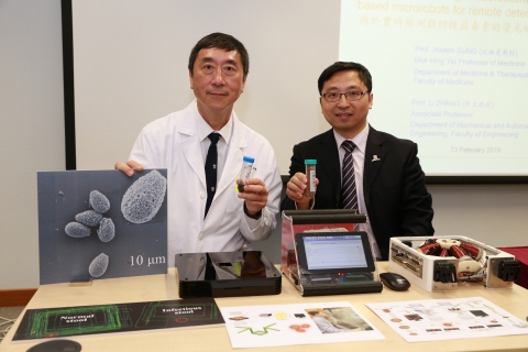
Microrobots have attracted much interest as an emerging medical tool which has great potential in surgery, therapy, imaging and diagnostics. A collaborative research team from the Faculty of Engineering and Faculty of Medicine has recently developed fungi spore-inspired microrobots to detect Clostridium difficile (C. difficile) bacterial toxins. The microrobots are active sensors capable of detecting toxins accurately within 15 minutes, based on a specific combination. The finding has been published in Science Advances, a scientific journal of the American Association for the Advancement of Science (AAAS). The team has also submitted a proposal to the Innovation and Technology Fund (ITF) on the development of a fully-automated, microrobots-based, quick diagnostics platform that can improve the efficiency and accuracy of clinical tests.
Early discovery of C. difficile patients helps infection control
C. difficile infection is the most common hospital acquired enteric infection. The toxins secreted by C. difficile will cause diarrhea, fever and hematochezia. In some cases, patients may develop life-threatening peritonitis and sepsis. The infection most commonly affects people who have recently been treated with antibiotics, while other major risk factors include old age, chronic illness and inflammatory bowel disease.
Studies conducted by Faculty of Medicine at CUHK suggest an increasing trend in the incidence of C. difficile infection which has placed a huge burden on the public health system in Hong Kong. The incidence of infection increased from 15.4 cases per 100,000 persons in 2006 to 36.3 cases per 100,000 persons in 2014. Over 10% of patients who have been hospitalised for two weeks are infected with C. difficile.
Currently, the stool samples of hospitalised patients with diarrhea are being tested in the laboratory to determine the presence of C. difficile. The process normally requires 1 to 2 days.
Professor Joseph SUNG, Mok Hing Yiu Professor of Medicine and Director of the Institute of Digestive Disease at CUHK, stated, “C. difficile spreads by contact with the excreta of infected persons or contaminated surfaces. Since the balance of healthy and pathogenic bacteria in the patient’s bowel has been damaged, it is hard to cure and easy to relapse. The development of a fast, accurate, simple and inexpensive test tool to shorten the diagnosis time allows doctors to give appropriate treatment and hospitals to carry out infection management measures in the earliest possible time, which can effectively prevent the spread of bacteria.”
The research team led by Professor Li ZHANG, Associate Professor, Department of Mechanical and Automation Engineering at CUHK, has developed fluorescent magnetic spore-based microrobots (FMSMs) to shorten the detection time. Each of these devices carry functionalised carbon dots that emit fluorescence, the intensity of which will gradually decrease during “on-the-fly” reaction with C. difficile toxins. The FMSMs unique and intricate three-dimensional architecture enables easy spreading and swarming in diluted stool samples. Such a continuous and efficient movement acts as active searching, thus facilitating higher detection efficiency and sensitivity than static counterparts. This enables the reaction even if the sample has a low concentration of toxins. Also, when applying an external magnetic field, FMSMs can perform a controllable movement in the stool samples and be tracked with automation in an easy manner.
Professor Zhang said, “In the experiment, all FMSMs placed into stool samples infected by C. difficile no longer emit fluorescence in just 15 minutes. This new motion-based detection technique provides a promising solution to the rapid clinical sensing to supplement, or potentially replace the current detection methods in clinic. This new technology eventually provides opportunities to develop a multiplex new quick-sensing system not only for C. difficile toxins, but also for many bioanalytical fields including food, chemicals and early diagnosis of other bacteria-infected diseases. Based on this novel microrobotic sensing probe, we will move forward to construct an automated microrobotic platform for practical diagnostic application that can be used in clinics and hospitals.”
CUHK committed to enhancing medical technology focusing in medical robotic
In recent years, the Hong Kong Government has been committed to promoting innovation and technology (I&T). These extremely exciting developments include the establishment of I&T clusters on healthcare technologies, artificial intelligence, and robotic technologies. CUHK is a leading institute for these areas in Asia. The establishment of the Chow Yuk Ho Technology Centre for Innovative Medicine aims to facilitate interdisciplinary collaboration between the Faculty of Engineering and the Faculty of Medicine, and to develop innovative technologies for clinical applications. Recently, CUHK has deepened ties with three leading international institutes for transdisciplinary medical robotics research to reshape the future of diagnosis and treatment.
Professor Zhang has devoted himself to the development of micro- and nano-robots, especially for medical use. Currently, he is leading his research team to improve the performance and functions of these micro-devices by paying close attention to their structural design, material properties, and control system.